


 Foundational characteristics of cancer include proliferation, angiogenesis, migration, evasion of apoptosis, and cellular immortality. Find key markers for these cellular processes and antibodies to detect them.
 The SUMOplot™ Analysis Program predicts and scores sumoylation sites in your protein. SUMOylation is a post-translational modification involved in various cellular processes, such as nuclear-cytosolic transport, transcriptional regulation, apoptosis, protein stability, response to stress, and progression through the cell cycle.
 The Autophagy Receptor Motif Plotter predicts and scores autophagy receptor binding sites in your protein. Identifying proteins connected to this pathway is critical to understanding the role of autophagy in physiological as well as pathological processes such as development, differentiation, neurodegenerative diseases, stress, infection, and cancer.
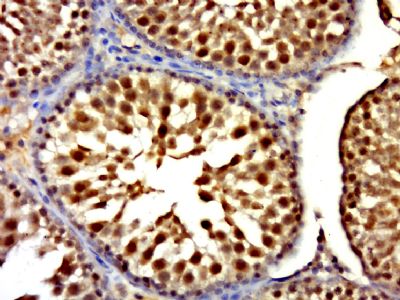
RNF6 Polyclonal Antibody
Purified Rabbit Polyclonal Antibody (Pab)
- SPECIFICATION
- CITATIONS
- PROTOCOLS
- BACKGROUND

Application
| WB, IHC-P, IHC-F, IF, ICC, E |
|---|---|
| Primary Accession | Q9Y252 |
| Reactivity | Rat, Dog, Bovine |
| Host | Rabbit |
| Clonality | Polyclonal |
| Calculated MW | 78091 Da |
| Gene ID | 6049 |
|---|---|
| Other Names | E3 ubiquitin-protein ligase RNF6, 2.3.2.27, RING-type E3 ubiquitin transferase RNF6, RNF6, SPG2 |
| Dilution | WB=1:500-2000,IHC-P=1:100-500,IHC-F=1:100-500,ICC=1:100-500,IF=1:50-200,ELISA=1:5000-10000 |
| Storage | Store at -20 ℃ for one year. Avoid repeated freeze/thaw cycles. When reconstituted in sterile pH 7.4 0.01M PBS or diluent of antibody the antibody is stable for at least two weeks at 2-4 ℃. |
| Name | RNF6 {ECO:0000303|PubMed:10331950, ECO:0000312|HGNC:HGNC:10069} |
|---|---|
| Function | E3 ubiquitin-protein ligase mediating 'Lys-48'-linked polyubiquitination of LIMK1 and its subsequent targeting to the proteasome for degradation (By similarity). Negatively regulates axonal outgrowth through regulation of the LIMK1 turnover (By similarity). Mediates 'Lys-6' and 'Lys-27'-linked polyubiquitination of AR/androgen receptor thereby modulating its transcriptional activity (PubMed:19345326). May also bind DNA and function as a transcriptional regulator (By similarity). Mediates polyubiquitination of QKI in macrophages, leading to its degradation (By similarity). |
| Cellular Location | Nucleus. Cytoplasm. Cell projection, axon {ECO:0000250|UniProtKB:Q9DBU5}. Nucleus, PML body {ECO:0000250|UniProtKB:Q9DBU5}. Note=Localizes to the PML nuclear bodies in Sertoli cells. {ECO:0000250|UniProtKB:Q9DBU5} |
| Tissue Location | Weakly expressed in peripheral blood, spleen, prostate, testis and ovary (PubMed:19345326). According to a report, it is preferentially expressed in testis and ovary and hardly detected in other tissues (PubMed:18368307). |
Research Areas
Citations (0)

Thousands of laboratories across the world have published research that depended on the performance of antibodies from Abcepta to advance their research. Check out links to articles that cite our products in major peer-reviewed journals, organized by research category.
Submit your citation using an Abcepta antibody to
info@abcepta.com, and receive a free "I Love Antibodies" mug.
info@abcepta.com, and receive a free "I Love Antibodies" mug.

Application Protocols
Provided below are standard protocols that you may find useful for product applications.
Abcepta welcomes feedback from its customers.
If you have used an Abcepta product and would like to share how it has performed, please click on the "Submit Review" button and provide the requested information. Our staff will examine and post your review and contact you if needed.
If you have any additional inquiries please email technical services at tech@abcepta.com.
$ 385.00
Cat# AP59170
Ordering Information
United States
AlbaniaAustraliaAustriaBelgiumBosnia & HerzegovinaBrazilBulgariaCanadaCentral AmericaChinaCroatiaCyprusCzech RepublicDenmarkEstoniaFinlandFranceGermanyGreeceHong KongHungaryIcelandIndiaIndonesiaIrelandIsraelItalyJapanLatviaLithuaniaLuxembourgMacedoniaMalaysiaMaltaMexicoNetherlandsNew ZealandNorwayPakistanPolandPortugalRomaniaSerbiaSingaporeSlovakiaSloveniaSouth AfricaSouth KoreaSpainSwedenSwitzerlandTaiwanTurkeyUnited KingdomUnited StatesVietnamWorldwideOthers
USA Headquarters
(888) 735-7227 / (858) 622-0099 or (858) 875-1900
Other Products
Shipping Information
Domestic orders (in stock items)
Shipped out the same day. Orders placed after 1 PM (PST) will ship out the next business day.
International orders
Contact your local distributors
