


 Foundational characteristics of cancer include proliferation, angiogenesis, migration, evasion of apoptosis, and cellular immortality. Find key markers for these cellular processes and antibodies to detect them.
 The SUMOplot™ Analysis Program predicts and scores sumoylation sites in your protein. SUMOylation is a post-translational modification involved in various cellular processes, such as nuclear-cytosolic transport, transcriptional regulation, apoptosis, protein stability, response to stress, and progression through the cell cycle.
 The Autophagy Receptor Motif Plotter predicts and scores autophagy receptor binding sites in your protein. Identifying proteins connected to this pathway is critical to understanding the role of autophagy in physiological as well as pathological processes such as development, differentiation, neurodegenerative diseases, stress, infection, and cancer.
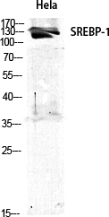
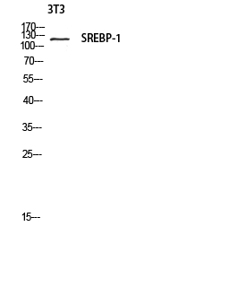
SREBP-1 Polyclonal Antibody
- SPECIFICATION
- CITATIONS
- PROTOCOLS
- BACKGROUND

Application
| WB, IHC-P |
|---|---|
| Primary Accession | P36956 |
| Reactivity | Human, Rat |
| Host | Rabbit |
| Clonality | Polyclonal |
| Calculated MW | 121675 Da |
| Gene ID | 6720 |
|---|---|
| Other Names | SREBF1; BHLHD1; SREBP1; Sterol regulatory element-binding protein 1; SREBP-1; Class D basic helix-loop-helix protein 1; bHLHd1; Sterol regulatory element-binding transcription factor 1 |
| Dilution | WB~~Western Blot: 1/500 - 1/2000. Immunohistochemistry: 1/100 - 1/300. ELISA: 1/20000. Not yet tested in other applications. IHC-P~~N/A |
| Format | Liquid in PBS containing 50% glycerol, 0.5% BSA and 0.09% (W/V) sodium azide. |
| Storage Conditions | -20℃ |
| Name | SREBF1 {ECO:0000303|PubMed:7759101, ECO:0000312|HGNC:HGNC:11289} |
|---|---|
| Function | [Sterol regulatory element-binding protein 1]: Precursor of the transcription factor form (Processed sterol regulatory element- binding protein 1), which is embedded in the endoplasmic reticulum membrane (PubMed:32322062). Low sterol concentrations promote processing of this form, releasing the transcription factor form that translocates into the nucleus and activates transcription of genes involved in cholesterol biosynthesis and lipid homeostasis (By similarity). |
| Cellular Location | [Sterol regulatory element-binding protein 1]: Endoplasmic reticulum membrane; Multi- pass membrane protein. Golgi apparatus membrane; Multi-pass membrane protein. Cytoplasmic vesicle, COPII-coated vesicle membrane {ECO:0000250|UniProtKB:Q9WTN3}; Multi-pass membrane protein. Note=At high sterol concentrations, the SCAP-SREBP is retained in the endoplasmic reticulum. Low sterol concentrations promote recruitment into COPII-coated vesicles and transport of the SCAP-SREBP to the Golgi, where it is processed {ECO:0000250|UniProtKB:Q9WTN3} [Isoform SREBP-1aDelta]: Nucleus |
| Tissue Location | Expressed in a wide variety of tissues, most abundant in liver and adrenal gland (PubMed:8402897). In fetal tissues lung and liver shows highest expression (PubMed:8402897) [Isoform SREBP-1C]: Predominantly expressed in liver and adipose tissues (PubMed:8402897). Also expressed in kidney, brain, white fat, and muscle (PubMed:8402897) |

Thousands of laboratories across the world have published research that depended on the performance of antibodies from Abcepta to advance their research. Check out links to articles that cite our products in major peer-reviewed journals, organized by research category.
info@abcepta.com, and receive a free "I Love Antibodies" mug.

Provided below are standard protocols that you may find useful for product applications.
Background
Transcriptional activator required for lipid homeostasis. Regulates transcription of the LDL receptor gene as well as the fatty acid and to a lesser degree the cholesterol synthesis pathway (By similarity). Binds to the sterol regulatory element 1 (SRE-1) (5'-ATCACCCCAC-3'). Has dual sequence specificity binding to both an E-box motif (5'-ATCACGTGA-3') and to SRE-1 (5'-ATCACCCCAC-3').
If you have used an Abcepta product and would like to share how it has performed, please click on the "Submit Review" button and provide the requested information. Our staff will examine and post your review and contact you if needed.
If you have any additional inquiries please email technical services at tech@abcepta.com.
Ordering Information
Other Products
Shipping Information
