


 Foundational characteristics of cancer include proliferation, angiogenesis, migration, evasion of apoptosis, and cellular immortality. Find key markers for these cellular processes and antibodies to detect them.
 The SUMOplot™ Analysis Program predicts and scores sumoylation sites in your protein. SUMOylation is a post-translational modification involved in various cellular processes, such as nuclear-cytosolic transport, transcriptional regulation, apoptosis, protein stability, response to stress, and progression through the cell cycle.
 The Autophagy Receptor Motif Plotter predicts and scores autophagy receptor binding sites in your protein. Identifying proteins connected to this pathway is critical to understanding the role of autophagy in physiological as well as pathological processes such as development, differentiation, neurodegenerative diseases, stress, infection, and cancer.
MAD1 Antibody
Rabbit mAb
- SPECIFICATION
- CITATIONS
- PROTOCOLS
- BACKGROUND

Application
| WB |
|---|---|
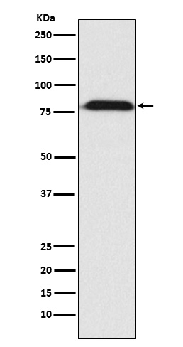
| Primary Accession | Q9Y6D9 |
| Reactivity | Rat |
| Clonality | Monoclonal |
| Other Names | hMAD1; HsMAD1; MAD1; MAD1L1; PIG9; TP53I9; TXBP181; |
| Isotype | Rabbit IgG |
| Host | Rabbit |
| Calculated MW | 83067 Da |
| Dilution | WB 1:500~1:2000 |
|---|---|
| Purification | Affinity-chromatography |
| Immunogen | A synthesized peptide derived from human MAD1 |
| Description | Component of the spindle-assembly checkpoint that prevents the onset of anaphase until all chromosomes are properly aligned at the metaphase plate. |
| Storage Condition and Buffer | Rabbit IgG in phosphate buffered saline , pH 7.4, 150mM NaCl, 0.02% sodium azide and 50% glycerol. Store at +4°C short term. Store at -20°C long term. Avoid freeze / thaw cycle. |
| Name | MAD1L1 |
|---|---|
| Synonyms | MAD1, TXBP181 |
| Function | Component of the spindle-assembly checkpoint that prevents the onset of anaphase until all chromosomes are properly aligned at the metaphase plate (PubMed:10049595, PubMed:20133940, PubMed:29162720). Forms a heterotetrameric complex with the closed conformation form of MAD2L1 (C-MAD2) at unattached kinetochores during prometaphase, recruits an open conformation of MAD2L1 (O-MAD2) and promotes the conversion of O-MAD2 to C-MAD2, which ensures mitotic checkpoint signaling (PubMed:29162720). |
| Cellular Location | Nucleus. Chromosome, centromere, kinetochore. Nucleus envelope Cytoplasm, cytoskeleton, microtubule organizing center, centrosome. Cytoplasm, cytoskeleton, spindle. Cytoplasm, cytoskeleton, spindle pole. Note=Co- localizes with TPR at the nucleus envelope during interphase and throughout the cell cycle (PubMed:18981471, PubMed:22351768). From the beginning to the end of mitosis, it is seen to move from a diffusely nuclear distribution to the centrosome, to the spindle midzone and finally to the midbody (PubMed:9546394). Localizes to kinetochores during prometaphase (PubMed:22351768, PubMed:29162720). Does not localize to kinetochores during metaphase (PubMed:29162720) Colocalizes with NEK2 at the kinetochore (PubMed:14978040). Colocalizes with IK at spindle poles during metaphase and anaphase (PubMed:22351768). |
| Tissue Location | [Isoform 1]: Expressed in hepatocellular carcinomas and hepatoma cell lines (at protein level) |
Research Areas
Citations (0)

Thousands of laboratories across the world have published research that depended on the performance of antibodies from Abcepta to advance their research. Check out links to articles that cite our products in major peer-reviewed journals, organized by research category.
Submit your citation using an Abcepta antibody to
info@abcepta.com, and receive a free "I Love Antibodies" mug.
info@abcepta.com, and receive a free "I Love Antibodies" mug.

Application Protocols
Provided below are standard protocols that you may find useful for product applications.
Abcepta welcomes feedback from its customers.
If you have used an Abcepta product and would like to share how it has performed, please click on the "Submit Review" button and provide the requested information. Our staff will examine and post your review and contact you if needed.
If you have any additional inquiries please email technical services at tech@abcepta.com.
$ 195.00
$ 385.00
Cat# AP92296
Ordering Information
United States
AlbaniaAustraliaAustriaBelgiumBosnia & HerzegovinaBrazilBulgariaCanadaCentral AmericaChinaCroatiaCyprusCzech RepublicDenmarkEstoniaFinlandFranceGermanyGreeceHong KongHungaryIcelandIndiaIndonesiaIrelandIsraelItalyJapanLatviaLithuaniaLuxembourgMacedoniaMalaysiaMaltaMexicoNetherlandsNew ZealandNorwayPakistanPolandPortugalRomaniaSerbiaSingaporeSlovakiaSloveniaSouth AfricaSouth KoreaSpainSwedenSwitzerlandTaiwanTurkeyUnited KingdomUnited StatesVietnamWorldwideOthers
USA Headquarters
(888) 735-7227 / (858) 622-0099 or (858) 875-1900
Other Products
Shipping Information
Domestic orders (in stock items)
Shipped out the same day. Orders placed after 1 PM (PST) will ship out the next business day.
International orders
Contact your local distributors
