


 Foundational characteristics of cancer include proliferation, angiogenesis, migration, evasion of apoptosis, and cellular immortality. Find key markers for these cellular processes and antibodies to detect them.
 The SUMOplot™ Analysis Program predicts and scores sumoylation sites in your protein. SUMOylation is a post-translational modification involved in various cellular processes, such as nuclear-cytosolic transport, transcriptional regulation, apoptosis, protein stability, response to stress, and progression through the cell cycle.
 The Autophagy Receptor Motif Plotter predicts and scores autophagy receptor binding sites in your protein. Identifying proteins connected to this pathway is critical to understanding the role of autophagy in physiological as well as pathological processes such as development, differentiation, neurodegenerative diseases, stress, infection, and cancer.
Bik Antibody
- SPECIFICATION
- CITATIONS
- PROTOCOLS
- BACKGROUND

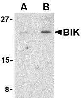
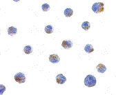
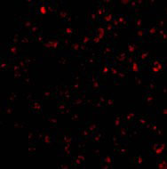
Application
| WB, IF, ICC, E |
|---|---|
| Primary Accession | Q13323 |
| Other Accession | CAG30276, 47678311 |
| Reactivity | Human, Mouse |
| Host | Rabbit |
| Clonality | Polyclonal |
| Isotype | IgG |
| Calculated MW | Predicted: 18 kDa Observed: 22 kDa |
| Application Notes | BIK antibody can be used for the detection of BIK by Western blot at 1 - 2 µg/mL. Antibody can also be used for immunocytochemistry starting at 1 µg/mL. For immunofluorescence start at 10 µg/mL. |
| Gene ID | 638 |
|---|---|
| Other Names | Bik Antibody: BP4, NBK, BIP1, Bcl-2-interacting killer, Apoptosis inducer NBK, BCL2-interacting killer (apoptosis-inducing) |
| Target/Specificity | BIK; |
| Reconstitution & Storage | Bik antibody can be stored at 4℃ for three months and -20℃, stable for up to one year. As with all antibodies care should be taken to avoid repeated freeze thaw cycles. Antibodies should not be exposed to prolonged high temperatures. |
| Precautions | Bik Antibody is for research use only and not for use in diagnostic or therapeutic procedures. |
| Name | BIK {ECO:0000303|PubMed:7478623, ECO:0000312|HGNC:HGNC:1051} |
|---|---|
| Function | Accelerates programmed cell death. Association to the apoptosis repressors Bcl-X(L), BHRF1, Bcl-2 or its adenovirus homolog E1B 19k protein suppresses this death-promoting activity. Does not interact with BAX. |
| Cellular Location | Endomembrane system; Single-pass membrane protein. Mitochondrion membrane {ECO:0000250|UniProtKB:O70337}; Single-pass membrane protein. Note=Around the nuclear envelope, and in cytoplasmic membranes. |

Thousands of laboratories across the world have published research that depended on the performance of antibodies from Abcepta to advance their research. Check out links to articles that cite our products in major peer-reviewed journals, organized by research category.
info@abcepta.com, and receive a free "I Love Antibodies" mug.

Provided below are standard protocols that you may find useful for product applications.
Background
Bik Antibody: Apoptosis plays a major role in normal organism development, tissue homeostasis, and removal of damaged cells and is caused by the activation of proteolytic enzymes termed caspases. Proteins that comprise the Bcl-2 family appear to control the activation of these enzymes. One such protein BIK was recently identified as an endoplasmic reticulum (ER)-residing pro-apoptotic member of the Bcl-2 homology domain-3 (BH3)-only group of the Bcl-2 family that stimulates mitochondrial release of cytochrome c following p53 induction of apoptosis. A significant fraction of BIK is found as an ER transmembrane protein, with most of the protein facing the cytosol. Restricting BIK to the ER membrane by replacing the transmembrane region with that of the ER-selective membrane anchor of cytochrome b resulted in a decreased cytochrome c release from mitochondria and a corresponding drop in cell death. Recent evidence suggests that BIK cooperates with NOXA, another BH3-only protein, to somehow enhance the activation of Bax to stimulate the rapid release of cytochrome c from mitochondria.
References
Lockshin RA, Osborne B, and Zakeri Z. Cell death in the third millennium. Cell Death Differ. 2000; 7:2-7.
Germain M, Mathai JP, and Shore GC. BH-3-only BIK functions at the endoplasmic reticulum to stimulate cytochrome c release from mitochondria. J. Biol. Chem. 277:18053-60.
Germain M, Mathai JP, McBride HM, et al. Endoplasmic reticulum BIK initiates DRP1-regulated remodelling of mitochondrial cristae during apoptosis. EMBO J. 2005; 24:1546-56.
If you have used an Abcepta product and would like to share how it has performed, please click on the "Submit Review" button and provide the requested information. Our staff will examine and post your review and contact you if needed.
If you have any additional inquiries please email technical services at tech@abcepta.com.
Ordering Information
Other Products
Shipping Information
