


 Foundational characteristics of cancer include proliferation, angiogenesis, migration, evasion of apoptosis, and cellular immortality. Find key markers for these cellular processes and antibodies to detect them.
 The SUMOplot™ Analysis Program predicts and scores sumoylation sites in your protein. SUMOylation is a post-translational modification involved in various cellular processes, such as nuclear-cytosolic transport, transcriptional regulation, apoptosis, protein stability, response to stress, and progression through the cell cycle.
 The Autophagy Receptor Motif Plotter predicts and scores autophagy receptor binding sites in your protein. Identifying proteins connected to this pathway is critical to understanding the role of autophagy in physiological as well as pathological processes such as development, differentiation, neurodegenerative diseases, stress, infection, and cancer.
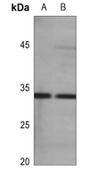
Anti-Aquaporin 3 Antibody
Rabbit polyclonal antibody to Aquaporin 3
- SPECIFICATION
- CITATIONS
- PROTOCOLS
- BACKGROUND

Application
| WB |
|---|---|
| Primary Accession | Q92482 |
| Other Accession | Q8R2N1 |
| Reactivity | Human, Mouse, Rat, Pig, Bovine |
| Host | Rabbit |
| Clonality | Polyclonal |
| Calculated MW | 31544 Da |
| Gene ID | 360 |
|---|---|
| Other Names | Aquaporin-3; AQP-3; Aquaglyceroporin-3 |
| Target/Specificity | KLH-conjugated synthetic peptide encompassing a sequence within the center region of human Aquaporin 3. The exact sequence is proprietary. |
| Dilution | WB~~WB (1/500 - 1/1000) |
| Format | Liquid in 0.42% Potassium phosphate, 0.87% Sodium chloride, pH 7.3, 30% glycerol, and 0.09% (W/V) sodium azide. |
| Storage | Store at -20 °C.Stable for 12 months from date of receipt |
| Name | AQP3 {ECO:0000303|PubMed:7558005, ECO:0000312|HGNC:HGNC:636} |
|---|---|
| Function | Aquaglyceroporins form homotetrameric transmembrane channels, with each monomer independently mediating glycerol and water transport across the plasma membrane along their osmotic gradient (PubMed:12239222, PubMed:30420639). Could also be permeable to urea (By similarity). Also participates in cell permeability to H2O2 and H2O2- mediated signaling (PubMed:20724658). In skin, transports glycerol to the epidermis and stratum corneum, where it maintains hydration, elasticity, and supports lipid biosynthesis for barrier repair (By similarity). In kidney, contributes to the reabsorption of water, helping the body maintain proper fluid balance (By similarity). |
| Cellular Location | Cell membrane; Multi-pass membrane protein {ECO:0000250|UniProtKB:O14520}. Basolateral cell membrane {ECO:0000250|UniProtKB:P47862}; Multi-pass membrane protein {ECO:0000250|UniProtKB:O14520} |
| Tissue Location | Widely expressed in epithelial cells of kidney (collecting ducts) and airways, in keratinocytes, immature dendritic cells and erythrocytes. Isoform 2 is not detectable in erythrocytes at the protein level |

Thousands of laboratories across the world have published research that depended on the performance of antibodies from Abcepta to advance their research. Check out links to articles that cite our products in major peer-reviewed journals, organized by research category.
info@abcepta.com, and receive a free "I Love Antibodies" mug.

Provided below are standard protocols that you may find useful for product applications.
Background
KLH-conjugated synthetic peptide encompassing a sequence within the center region of human Aquaporin 3. The exact sequence is proprietary.
If you have used an Abcepta product and would like to share how it has performed, please click on the "Submit Review" button and provide the requested information. Our staff will examine and post your review and contact you if needed.
If you have any additional inquiries please email technical services at tech@abcepta.com.
Ordering Information
Other Products
Shipping Information
